Welcome to visitShanghai Chinon medical Model & Equipment Manufacturing Co., LTD
In medical education, pelvic anatomy has always been one of the basic contents of learning the structure and function of the human body. For medical students, understanding the anatomy of the pelvis is not only vital to the study of urology, obstetrics and gynecology, surgery and other disciplines, but also has a direct impact on the success rate of clinical operations and operations. Traditional teaching methods, such as anatomical diagrams, 2D images, and cadaver anatomy practice, have certain educational significance, but often fail to fully demonstrate the complexity of three-dimensional structures. Can the male pelvis model, as a modern teaching tool, improve the learning effect of pelvic anatomy? This issue has received wide attention in recent years
1. Visual display of three-dimensional structure

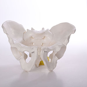
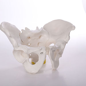
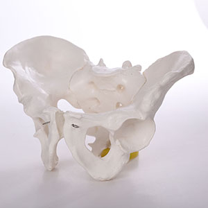
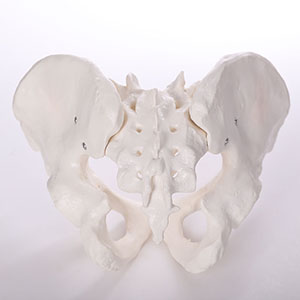
Male pelvic models, especially those using 3D printing technology, can clearly reproduce the three-dimensional structure of the pelvis, and can be disassembled, rotated, and adjusted. This highly reductive structure allows students to view the constituent parts of the pelvis, such as the pelvis, hip bones, sacrococcygeal end and their relationship to other organs from all directions, resulting in a more intuitive understanding of the complex anatomy of the pelvis. Compared with traditional flat images or 2D videos, 3D models provide students with a more vivid and three-dimensional learning experience.
According to a 2019 study published in Medical Education Online, students who used 3D models to teach pelvic anatomy scored, on average, about 18 percent better on their anatomy knowledge on final exams than students who were taught in the traditional way. In addition, students have a more comprehensive understanding of the pelvic structure and are able to accurately identify the various anatomical markers.
2. Enhance students' spatial cognition ability

Learning pelvic anatomy not only requires students to memorize the name and location of each organ, but also requires them to have strong spatial cognition ability. Through the male pelvis model, students are able to perceive the relative position of the various parts of the pelvis in three dimensions by touching, rotating and disassembling, which helps to strengthen the connection between spatial memory and anatomical structure.
An experimental study published in 2020 in Advances in Health Sciences Education found that students who used a three-dimensional pelvis model showed significant improvements in their spatial cognition compared to students who learned from a traditional two-dimensional textbook. Through interactive manipulation, students are able to more accurately locate the lesion site or anatomical structure in practical clinical applications, thereby improving the accuracy of diagnosis and treatment.
3. Improve the sense of participation and interaction in learning

The use of the male pelvis model is not only a process of viewing and memorizing, but also a process of interactive learning. Students can directly operate the model, carry out structural comparison and practical operation, and enhance the sense of participation in learning. Through simulated surgery, manual disassembly and reconstruction of the various components of the pelvis, students can not only deepen their understanding of theoretical knowledge, but also develop surgical skills and clinical judgment in practice.
In 2021, a study in the Journal of Surgical Education showed that students who participated in the teaching of 3D pelvic models had higher interaction and engagement in mastering basic anatomy and clinical skills than students who simply went through lectures and reading materials. According to the data, 90% of the students believe that they have a better understanding of the pelvis and improved their ability to operate it after using the 3D pelvis model.
4. Personalized learning and self-assessment

The male pelvis model not only helps in classroom teaching, but also provides conditions for personalized learning. Students can operate the model repeatedly according to their own learning progress, and carry out self-assessment and knowledge review at any time. For some of the more difficult anatomical structures, students can enhance their memory through repeated practice without being limited by time and space.
For example, the 3D pelvis model allows students to disassemble and reassemble the pelvis structure at home or in the study room, which helps students to systematically review before the exam and make up for the shortcomings of classroom learning. A 2020 study in BMC Medical Education found that students who used 3D models for revision generally improved their test scores, especially in complex and confusing areas such as the hip bone and sacrum.
5. Summary and outlook
In conclusion, the application of male pelvis model in medical education can significantly improve the effect of pelvic anatomy learning. Through the visual display of three-dimensional structure, enhancing spatial cognitive ability, promoting interactive learning and personalized review, the model not only improves students' understanding of pelvic structure, but also plays a positive role in practical operation and clinical application ability. With the continuous progress of 3D printing and virtual reality technology, the future male pelvis model is expected to be more intelligent and personalized, and become an indispensable tool in medical education.
For medical students, the use of this model can not only help them achieve better results in theoretical learning, but also lay a solid foundation for future clinical practice, thus promoting medical education to a more efficient and accurate direction.
|
NEXTпјљProstate test model: Can it help doctors improve test accuracy?
LASTпјљCan pediatric enema models help reduce care errors? |
Return list |